| Home Farrier Wisdom Medical Index | First Posted: July 15, 2008 Updated: Jan 21, 2020 | |
Abscesses, Cracks, Sole Bruises and Corns in the Horseby Debora JohnsonWhat Is An Abscess?
The Merck Vet Manual defines a foot abscess as: "A foot abscess is a necrotizing or purulent infection involving the distal interphalangeal tissues and sometimes the joint. The incidence is usually sporadic, but up to 15% of the flock may be affected. The 2 organisms most consistently recovered from foot abscess are Fusobacterium necrophorum and Arcanobacterium (Actinomyces) pyogenes . Most commonly, foot abscesses develop as a complication of ovine interdigital dermatitis (see Ovine Interdigital Dermatitis) by extension of the necrotic process into the subcutis and then into the distal interphalangeal joint. This joint is vulnerable to infection on the interdigital aspect where the joint capsule protrudes above the coronary border as the dorsal and volar pouches. At these 2 sites, the joint capsule is protected only by the interdigital skin and a minimal amount of subcutaneous tissue. Sporadic cases of foot abscess may also develop after penetration by sharp objects (eg, crusted snow, the stiff stubble of cut alfalfa fields) or careless paring of the hoof. Foot abscesses develop most often when the soil and pastures are wet. The disease causes an acute lameness that is usually restricted to one foot. In the early stages, it may be possible to express necrotic material through an opening in the interdigital skin via the channel caused by the bacterial invasion. Later, the sinuses may extend to break out at one or more points above the coronet. In ~50% of the cases, movement of the affected digit is exaggerated, which indicates that the ligaments about the distal interphalangeal joint have ruptured; displacement of the digit during locomotion and permanent deformity are likely. Acute lameness, swelling of one digit, and discharging sinuses distinguish foot abscess from footrot. Radiographs can help determine the extent of joint damage. Early treatment with parenteral antibiotics is often effective and may prevent joint infection. Once the infection becomes established in the joint, treatment is of limited value. Therapy should be aimed at maintaining the integrity of the joint ligaments by draining the abscess, bandaging to reduce stress on the ligaments, and countering the bacterial infection with antibiotics or sulfonamides. Although the prognosis for complete recovery is poor, in most cases the foot heals sufficiently to allow adequate locomotion after ~2 mo. Control depends on early treatment and avoidance of the conditions that lead to ovine interdigital dermatitis. Although F necrophorum vaccines are available, they have not proved to be entirely satisfactory." To simplify this definition as it relates to foot abscesses in the horse one could say an abscess is an infection. It involves the sensitive tissues of the horse's foot and may cause varying degrees of lameness. Sometimes you will hear the word "seedy toe" or "white line disease" used in relation to abscesses. Punctures can cause abscesses, too. Abscesses often migrate up the hoof and break out at the coronary band. This is usually the path of least resistance. After the abscess begins to drain the horse will usually show improvement. Symptoms of an abscess of the foot are lameness, increased pulse rate in the foot, and heat in the hoof. Hoof testers are helpful in locating the area of the abscess. Farriers and vets often pare away the sole area until they find the pus pocket. As the pus pocket begins to drain the abscess is flushed out with a solution that will be recommended by your vet or farrier. Then a germicide is used to irrigate the abscess and a drawing agent is used on cotton, for example, by packing the soaked cotton into the pared area. Ichthammol is a drawing agent often used. Sometimes soaking the horse's foot with epsom salts is necessary to draw out the abscess. Make sure not to get carried away paring out the sole area. The larger the hole in the sole the longer it takes to get your horse back to work. If the abscess is really severe antibiotics may be necessary.
Hoof Cracks
Sole Bruises
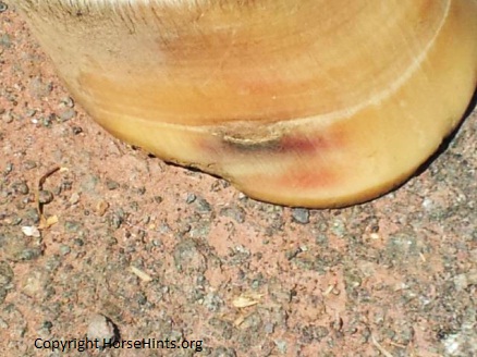
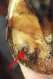
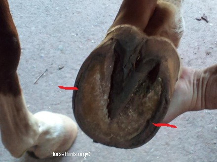 The image on the left has a bruise that shows red on the outside of the hoof. You can also see the flaring toward the bottom left and a "notch" out of the left side of the hoof. This horse also has "white line disease" if you pick up the foot and the beginning of "shelly feet."
Have you ever noticed red spots or specks on the sole, frog, white line or hoof of your horse's foot? Most likely these are bruises caused by trauma of some sort. The trauma could be a bruise from stepping on a rock or some other object. A horse that forges sometimes catches the soles of his own feet. It is important to have some hoof wall and not trim to short. My two horses are barefoot. Our farrier is very careful to leave enough hoof so that the soles do not get bruised on uneven ground. Don is truly wonder when it comes to shoeing. Sometimes, right after a trim, the horses will be "ouchy" for a day or two. We try to keep them on soft or grassy ground and off any rocks. We also use Keratox on their hooves to harden up the hoof and sole of the foot. I call it "liquid farrier." Although expensive, we think it is really an excellent product. The Merck Vet Manual says: "Bruising on the volar surface of the foot usually is caused by direct injury from stones, irregular ground, or other trauma. Poor shoeing, especially in horses with flat feet or dropped soles, predisposes to bruising, usually around the periphery of the sole. Bruising may or may not be associated with lameness, but if it becomes chronic, the affected area can become infected. Persistent, nonresponsive bruised sole suggests pedal osteitis."
Corns
"Corns are described as dry when only mild inflammatory changes exist, as moist when there is excess inflammatory exudate, and as suppurative once they become secondarily infected. When the foot is raised and the solar surface freed of dirt and loose horn, a discoloration, either red or reddish yellow, is noted. Supporting-leg lameness is an early sign, but lameness is not always seen. Tapping with a light hammer over the area or applying pressure with a hoof tester usually causes discomfort. If infection is present, pain is pronounced when pressure is applied with hoof testers; if not promptly treated, a tract may extend through to the coronet to produce a suppurating sinus.
The prognosis is favorable. In uncomplicated dry corns, relief from pressure on the affected area is the first consideration. This can be achieved by shortening the toe if it is too long and by applying a bar shoe to promote frog pressure. A three-quarter-bar shoe may be of value in relieving pressure. If the corn is suppurating, it should be drained immediately by a surgical opening directly through the sole. After drainage, the foot should be dressed to permit drainage. Hot foot baths and poultices may be helpful. The horse should be kept in a dry, clean box stall. After infection is controlled, the cavity can be packed with sterile gauze and topical antibiotic ointment, and a metal, rubber, or leather sole placed between the shoe and the foot. Parenteral antibacterial therapy is of questionable value unless systemic illness is present." Merck Vet Manual-Corns Simply: "Corns can be classified as dry or moist. Corns start out as bruises of the sensitive sole in the angle formed by the hoof wall and bars. This angle, where the wall and bars meet, is the seat area where corns originate. A dry corn is a red bruise in the seat of the corn area. The redness is caused by the horn tubular filling with blood from a ruptured vessel. A moist corn is yellow, with serum present. Corns are caused by unequal pressure and concussion created by a conformational fault or faulty trimming. Corns can be caused by overtrimming the heels, heel calks (heel shoes that have small cleats on the end of the shoes), short-heeled shoes, un level shoes (shoes that have not been leveled properly after being shaped to fit the hoof but are simply nailed on the hoof), or leaving the shoes on too long.
Corns can be prevented by eliminating the causes. Pressure on the corn seat also may be relieved by trimming the sole between the bars and the hoof wall so that it is 1/8 inch lower than the wall." For More Information: Horse Foot Bruises |