Horse Facts and Tips Medical Index Home | First Posted: Oct 13, 2012 Jan 21, 2020 | |
Eye Exams in Horsesby Debora JohnsonWhen it comes to the horse's eyes I have been told all my life that time is really important. Whether it is true or not--call the vet ASAP! With our horses we always call the vet right away, hopefully we will discover an eye problem within 24 hours. The horses are checked at least twice a day in the morning and in the evening. My husband and I routinely check each horse for the following:
Simple Diagnostic Tests Flourescein dye strips - Fluorescein dye is a water soluble dye which is unable to penetrate the lipid containing corneal or conjunctival eipthelium unless there is a break in its integrity. This allows the identification of damage in these structures which otherwise might be difficult to detect. Uses: All cases with signs of corneal disease and/or ocular pain especially ulceration, as part of complete ophthalmologic examination, check patency of naso-lacrimal drainage apparatus. Seidel's test: The Seidel Test is used to assess the presence of anterior chamber leakage in the cornea. It is used as a screening test for many corneal disorders including corneal post-trauma, corneal perforation and corneal degeneration. The Seidel Test is named after the German Ophthalmologist Erich Seidel (1882-1948). A fluorescein strip containing 10% fluorescein is applied topically to the affected area and is examined with a cobalt blue filter. At this point, the fluorescein appears green in color. Any changes in color or surface of the fluorescence area indicate the presence of corneal leakage. If the fluorescein strip turns pale upon application to the corneal surface, the test is positive for the corneal deformity. The change in the color of the fluorescein strip is due to dilution of fluorescein caused by the aqueous leakage in the cornea. Rose Bengal stain - To stain the cornea, Rose Bengal and/or Fluorescein strips. Fluorescein strips are more commonly used, and will stain areas where there is loss of epithelium. Rose Bengal will stain areas with devitalized epithelium, as well as reveal where epithelium is lost. Either strip should be held against the conjunctiva, or alternatively, the strip can be wetted with sterile saline and then the solution can be dropped across the eye. As the strip can be somewhat irritating if placed directly on the cornea, direct corneal contact should be avoided. If one suspects decreased tear production, a Schirmer tear test Corneal scrapings--Sometimes corneal scrapings are taken when ulcers are present. It is bacteria or fungus that is being targeted for treatment. Intraocular pressure - Although primary glaucoma exists in the horse, the secondary form appears to be most common. Trauma, lens luxation, and particularly uveitis are known causes of secondary glaucoma in the horse. Tono-Pens are often used for the test. Sometimes motor nerve blocks will be used. Ocular ultrasound is also used as an advanced technology for testing for glaucoma in the horse. 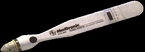 Tanopen Ultrasound of a horse's eye by John Kaufman DVM Exam Results After a complete eye examination has been performed on your horse the vet would be able to identify most of the following:
For More Information: Ophthalmic Exam/University of PA/VeterinaryEye Diseases Associated With Specific Horse Breeds Equine Opthomology/49th Ocala Equine conference Equine Recurrent Uveitis Equine Parasites Horse Vision Neurological Problem Signs in Horses |