If you cannot see images at all on my site click here for an explanation
Home
Medical Index
First Posted: Dec 30, 2009
Jan 21, 2020

Leg Problems Most Prevalent in Horses: Bone Spavin, Bog Spavin, Bowed Tendons, Tendon Sheath Effusion, Capped Hocks, Capped Knees, Knee Spavin, Osselets, Shin Splints or Bucked Shins, Speedy Cut (aka brush, cut down, grab a quarter), Sprained Suspensory Ligament, Stocking, Thoroughpin Bursal, Wind Puffs or Wind Galls

When does a horse begin to develop osteoarthritis? Typically the onset of osteoarthritis (OA) in adult horses is 4 to 6 years old, but that can vary a great deal due to breed of the horse and its use. Conformation is also a very important consideration leading to OA. A poorly conformed horse is more likely to be predisposed to an arthritic condition that would affect them earlier in life. Predisposing radiographic factors that are present in a young horse can also indicate possible future issues. Age for Onset of Osteoarthritis

Bone Spavin - Bone spavin is a bony growth within the lower hock joint. It is caused by osteoarthritis. The degree of lameness that results can be serious enough to end a horse's competitive career. Bone spavin is osteoarthritis, or the final phase of degenerative joint disease (DJD), in the lower three hock joints. It usually affects the two lowest joints of the hock (the tarsometatarsal and the distal intertarsal joints), with the third joint, the proximal intertarsal, being the least likely to develop bone spavin. This condition has more than one 'type.' Jack spavin is the term used if the lesion on the tarsal and carpal bones is large. If the pathology occurs higher in the joint than is typical, it is termed high spavin. Conversely, occult spavin does not produce any significant exostoses on the small tarsals, while bog spavin and blood spavin do not involve bony changes.
  
Causes:
  • Stress on the joints
  • quick and sliding stops such as those which occur during roping and reining. Mineral deficiencies Poor confirmation.
  • Concussion on hard surfaces such as cement, hard ground, etc.
  • Quick stops and turns used in Western competitions such as roping, team penning, etc.
  • Conformation problems
  • Deficiencies in diet. Lack of minerals. Poor nutrition.
  • Osteoarthritis
  • Degenerative joint disease
Bog Spavin - Bog Spavin is a swelling of the tibiotarsal joint of the horse's hock which, in itself, does not cause lameness. The joint becomes distended by excess synovial fluid and/or thickened synovial tissue bringing about a soft, fluctuant swelling on the front of the joint, as well as in the medial and lateral plantar pouches. Bog spavin is generally an indication of underlying pathology within the joint.
Causes:
  • Trauma to the elbow joint
  • Work on hard surfaces can cause wear and tear
  • Conformation problems
  • Nutritional deficiencies while young and growing
  • Defect in the tibiotarsal joint (the tibiotarsal joint (also known as the tarsocrural joint)
  • Synovitis - Inflammation of the lining of the joint capsule
  • Degenerative joint disease
  • Excessive strain of the joint capsule
  • In fully-mature horses, it is most likely because of chronic strain of the joint capsule

Bowed Tendons - Inflammation and in severe cases even rupture of the sheath encasing the tendon from the knee to the fetlock. One or both the deep flexor tendon and superficial flexor tendons on one and/or both front legs may be affected.

Pool House Veterinary Group and Equine Clinic
Causes:
  • Poor conformation: long, sloping pasterns and a long-toe, low-heel shape to the hoof (seen commonly in Thoroughbreds) predisposes a horse to tendinitis
  • Poor trimming and shoeing: such as a farrier that causes a hoof shape that predisposed the horse to tendon injuries (such as a long-toe and low-heel), or one that shoes a horse with toe grabs, which artificially create a long-toe and low heel by lifting the toe up.
  • Improper conditioning: such as working a horse at an intensity that it has not yet been conditioned for, working an unfit horse, and continuing to work an extremely fatigued horse.
  • Poor footing: working a horse on uneven or slippery footing can cause tendon strain, as well as deep, "thick" footing.
  • Each of these factors encourage the overextension of the fetlock and knee during work. Several of these factors at once can add up.
  • Direct trauma to a tendon: such as when a horse hits its front leg with a hind hoof.

Tendon Sheath Effusion - What is tendon sheath effusion?


Tendons attach muscles and bone, and are classified as flexors (flex a joint) or extensors (extend a joint). However, some tendons will flex multiple joints and extend another (the flexor tendons of the hind limb, for example, will flex the fetlock, pastern, and coffin joint, but extend the hock joint). In this case, they are classified according to whether they flex or extend the joints of the digit.

The following tendons are the main tendons found in the lower leg. When they pass over a joint, they are protected in a tendon sheath, which contains synovial fluid as a lubricant:

  • Common digital extensor: the common digital extensor muscle becomes tendon at the bottom third of the radius and continues down the front of the leg. The tendon pulls upward to extends the carpal, pastern, and coffin joints. It is the major extensor tendon of the leg. However, unlike the flexor tendons, a horse with a damaged or non-functional "extensor unit" (i.e., tendon and musculature) is not lame, but rapidly learns to compensate by "flicking" the lower limb using the carpal or tarsal extensor units. This seemed to describe what was happening with A Patchy's symptoms.
  • Image: Copyright HorseHints.org
    Common digital extensor tendon bottom third of the radius

  • Lateral digital extensor: the lateral digital extensor muscle becomes the lateral digital extensor tendon at the proximal portion of the metacarpus. The tendon continues down the front of the leg and inserts into the proximal portion of the first phalanx. Important in the treatment of stringhalt in the hindlimb. Extends the carpal, pastern, and coffin joints.
  • Deep digital flexor: 3 tendons of the deep digital flexor muscle travel distally and join at the carpus, were they pass through the carpal canal, and travel distally along the back of the leg, finally inserting into the palmar side of the third phalanx. Below the knee/hock, the tendon is superficial to the suspensory ligament, but deep to the SDFT. Fairly commonly injured by horses doing fast work, the DDFT is round in cross section.
  • Superficial digital flexor: Runs down the back of the leg, behind the carpus and cannon, branches below the fetlock and inserts into the distal side of the 1st phalanx and proximal side of the 2nd phalanx. Flexes the elbow, carpus and lower joints. Additionally, the superior check ligament inserts into this tendon from the caudal side of the radius. The SDFT is the most commonly injured tendon, and appears oval or flattened in cross section.

Capped Hocks (Bursal Enlargement) - up to the size of a tennis ball on the point of the hock. Horses may suffer from "capped hock," which is caused by the creation of a false bursa, a synovial sac beneath the skin. Capped hock is usually caused by trauma such as kicking or slipping when attempting to stand. In the absence of a wound, it does not require immediate veterinary attention and is usually only of cosmetic significance. On the other hand, a wound into the calcanean bursa is a serious problem. A capped hock is extremely unlikely to be a cause of lameness, even if severe.

Causes:
  • Trauma
  • A blow or bang to the point of the hock usually from horse kicking a wall or from lying down on a hard concrete surface.

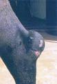
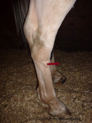
Capped Knees - An acute inflammation (bursal enlargement) or bruise of knee joint (carpitis) and/or the tendon that runs over the front of the knee.

Causes:
  • Blows to the knee joint (kicks, falls, injury on objects, etc.)
  • Joint stress
  • Rubbing
  • Excessive work
  • Poor conformation

Joint Capsule Damage: This actually happened to one of our horses. He fell on the ice and exposed the joint. We called the vet immediately and it turned out that the synovial fluid was leaking out into the surrounding tissues. The vet saved the day!

Curbs - A "curb" is a swelling at the back of the hock, traditionally considered to be due to a strain of the (long) plantar ligament (PL). The plantar ligament is a tough band of tissue that runs down behind the hock and helps maintain its stability. Inflammation on the upper rear of the cannon area just below the point of the hock may be seen.

Causes: Some horses may be susceptible to curbs because of their conformation (sickle hocks); others because of the type of work they do (jumping, racing Standardbreds or pulling loads.) Inflammation on the upper rear of the cannon area just below the point of the hock Caused by: Bang or blow the back of the leg - possibly from kicking the wall. Stress and trauma from a violent extension of the Plantar Ligament.
  • Poor conformation (sickle hocks)
  • Use: jumping, racing or pulling loads
  • Trauma to the back of the leg
  • Stall or wall kicking
  • Stress, trauma or over extension of the Plantar Ligament.

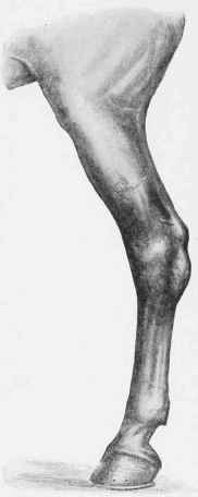
Knee Spavin - A weakening inflammation or growth on either the back or inside edge of a horse's hock. Bony growth at back of knee on inner side. Not very common. Carpal Spavin Surgery, Fine-Tuned

Causes:
  • Joint stress
  • Trauma caused by a blow of some sort

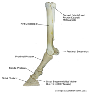
Osselets - A bony growth on the fetlock joint (the ankle of a horse; the joint located between the cannon bone and pastern bone) which causes inflammation of the surrounding tissues and membranes. There are other related terms such as arthritis, fetlock joint (the ankle of a horse; the joint located between the cannon bone and pastern bone), greenobsoletet (arthritis in the fetlock joint of young horses), osteoarthritis (a severe permanent form of arthritis that grows progressively worse in horses),synoviall fluidcorseletss (the lubricating fluid found in the joints of horses, and chronicobsoletet (the build up of excessive synovial fluid in a damaged joint, usually associated with osteoarthritis).

Causes:
  • Stress
  • Trauma
  • Overwork
  • Late stage development of "greencorseletss" that were not allowed to heal
  • Predisposition (genetics)
  • Inferior conformation

Inflammation of the bone above and at the back of the fetlock joint. Any of the ligaments or other anatomical structures of the joint, like theseamedd bones, may be involved. These problems may be particularly difficult to treat. Many horses have been left unsound as a consequence.


Sesamoid Injuries: Diagnosis, Treatment, and Prevention
Causes:
  • Trauma of some sort
  • Bang, blow, kick, rub, fall that injures the back of the fetlock joint

Shin Splints or Bucked Shins - Inflammation of the membrane that covers the Shin bone (cannon bone). A condition in which the splint bones contain excess calcification causing a bump or growth, usually as a result of improper healing of a fracture on the splint bones or due to irritation of the bones.

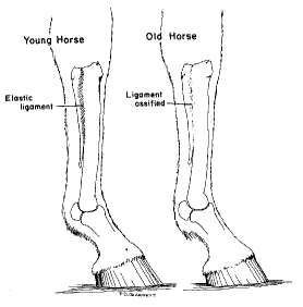
On each side of the cannon bone is a small bone known as the splint bone. The small splint bones are thin and taper to become a small knob about two-thirds of the way down the cannon bone. A ligament, located between the cannon bone and the splint bones, is quite elastic in young horses. As the horse ages, the ligament ossifies; that is, the ligament is replaced by bone and the three bones fuse. During ossification, there may be inflammation and pain. Jumping, running and working a horse during this time produces further irritation.

Splints usually occur in horses 2 to 5 years old. Most often it is the forelimbs which are affected. Splints rarely occur in the hind limbs. In older horses, the splint bones are fused solidly to the cannon bone.

The majority of splint problems occur on the medial side (inside) of the forelimbs. The medial splint bone usually is the one affected because it has a flat surface next to the knee. The lateral (outer) splint bone has a more slanted surface. When the weight is transmitted to these bones, the medial splint bone probably bears more weight than the lateral splint bone. Therefore, the ligament between the medial splint bone and the cannon bone is subjected to more stress than the outer ligament.

Lameness due to splints is most common in 2-year-old horses undergoing training. The lameness is most obvious while the horse is trotting or working or soon thereafter. Lameness may come and go or be present continuously for as long as a year. If you probe up and down along the cannon bone, the horse will flinch when the portion of the ligament undergoing ossification is touched. A large swelling or a number of smaller swellings due to ossification may occur along the length of the splint bones. After the ligament has ossified, the swelling and soreness usually disappears.

Causes:
  • Concussion
  • Working young horses too early
  • Rapid growth
  • Leg interference by striking the leg with a hoof

Speedy Cut - An injury to the hock caused by the over reaching gait of a rear leg. Related terms may be brush, cut down, grab a quarter. This is a common occurrence in horse racing.

Causes:
  • Bad conformation (brushing, forging, limb interference of any sort by self or something else)
  • Poor shoeing
  • Quick movements due to turnout, exercise, play, etc.

Sprained Ankle - Affecting one or more of the ligaments the support the fetlock joint.

Causess:
  • Stress
  • Trauma
  • Over extension such as forging, striking, etc.

Sprained Suspensory Ligament - The suspensory ligament is attached to the back of the upper cannon and knee (in the front legs) or hock (in the hind legs), runs downwards close to the back of the cannon and divides into two branches each of which attaches to a sesamoid bone, at the back of the fetlock, before ending attached to the upper pastern. The suspensory ligament supports the fetlock and protects it from hyperextension (i.e. dropping too low) at exercise.

Causes:
  • Stress
  • Trauma
  • Fatigue
  • Over extension

Stocking Up - Fluid retention in the lower limbs.

Causes:
  • Confinement
  • Lack of exercise
  • Circulatory problems
  • Too much rich food such as grain
  • Could be kidney problems
  • Predisposition (genetics)

Ligaments of the legs include:

  • Suspensory ligament: runs from the back of the cannon bone (between the two spint bones), then splits into two branches and attaches to the sesamoid bones at the bottom of the fetlock. Branches continue downward and attach to the extensor tendons. The main purpose of the suspensory is to support the fetlock joint, preventing it from overextending. Injury to this ligament is an important cause of lameness in performance horses. The suspensory is a modified muscle, the equine equivalent of the interosseous muscle, which contains both tendon fibers and residual muscle fibers.
  • Interosseous ligaments: connect the cannon bone to each splint bone. Injury to this ligament produces the condition known as splints.
  • Proximal and distal check ligaments: The proximal check ligament originates from the radius and attaches to the superficial digital flexor tendon. The distal check originates from the palmar carpal ligament and attaches to the deep digital flexor tendon, approximately 2/3-way down the metacarpus.
  • Plantar ligament: in the hind leg, runs down the lateral side of the tarsus, attaches to the fibular, 4th tarsal, and 3rd metatarsal bones. Injury leads to a condition known as curb.
  • Inter-sesamoidean ligaments: supporting ligaments, run between the two sesamoid bones.
  • Distal sesamoidean ligaments: run from the sesamoid bones to the two pastern bones. Important in the stay apparatus.
  • Impar ligament: runs between the navicular bone and the 3rd phalanx.
  • Annular ligament: goes around the back of the fetlock, surrounding the flexor tendons and their tendon sheath, attaching to the sesamoid bones. It helps to support the fetlock, and provides an enclosed "pulley" for the flexor tendons to run through.
  • Sacrosciatic ligament: Originates from the sacrum and coccygeal vertebrae, inserts into the pelvis.

Thoroughpin Bursal - "Thoroughpin is a distention of the tarsal sheath of the deep digital flexor tendon just above the hock. It is characterized by plantar fluid-filled swellings visible on both medial and lateral sides proximal to the tibiotarsal joint, which distinguish it from bog spavin ( Serous Tarsitis). It is usually unilateral and varies in size. The lesion is referred to as a tenosynovitis of traumatic origin, but it may not be associated with any detectable inflammation, pain, or lameness. It essentially constitutes a blemish and so is of major clinical importance in show horses. Treatment is by withdrawal of the fluid and injection of hyaluronic acid or a long-acting corticosteroid, which may need to be repeated until the swelling does not recur. Radiation therapy also helps reduce the secretory property of the tendon sheath." Merck Veterinary Manual

Causes:
  • Stress on tendon
  • Over extension
  • Trauma
  • Conformation problems of the hock joint

Wind Puffs or Wind Galls Soft - Puffiness in the area of the horse's ankle. The fetlock joint capsule and the flexor tendon sheath may retain fluid: "Spongy" swellings around the back, front and or side of the fetlock joint. The inflamed joint capsule distends with additional synovial fluid in an effort to protect against injury.
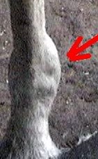
Causes:
  • Age
  • Concussion in the joint
  • Conformation/Genetics - Horses with coarse, meaty legs, round bones, poorly defined joints and veins are more prone to wind puffs.
  • Excessive exercise
  • Excessive load
  • Excessive work on hard surface
  • Fatigue
  • Hard work
  • Injury- Even in turnout!
  • Stress
  • Trailering a lot
  • Too much work at a young age before joints are developed
  • For More Information:

    Equine Lameness
    Lameness Gaited Horse
    Carpal Spavin in Horses - Bone Spavin
    Horse Bone Spurs
    Unsoundnesses and Blemishes of Horses: Feet and Legs
    Horse Tendons and Ligaments
    Tendon Sheath Effusion, Tenosynovitis
    The Conservative Approach for Healing Horses

    Home
    Medical Index