| Home Medical Index | First Posted: Apr 18, 2012 Jan 21, 2020 | |
Tendon Sheath Effusion or Tenosynovitisby Debora Johnson
Bill and I returned home from South America eager to see the horses. After rejoining the human race--a long flight--we went to the barn. The horses were so happy to see us. They came running to the fence from the bottom pasture to meet us--nickering all the way. Everything seemed fine. We groomed them and checked them all over but did not ride that day. Several days later we went out to ride. I noticed that my horse, A Patchy, was shaking his head back and forth as well as up and down--but only intermittently. There did not seem to be any annoying insects. He wears a bitless bridle so it was not a bit hurting him. His gait was not off and he wanted to go forward, but something just did not seem right. He never exhibited this behavior in the past. We went back to the barn and checked him all over for heat and swelling, looked in his ears for ticks, in his mouth for any problems, felt his hooves for heat, took the pulse in his legs, looked all around the bottom of his feet, no reaction from pressure testers on the bottom of his hoof, no foreign objects, etc. His appetite was normal. He was not dehydrated. We could not find anything. We went out again a few days later to ride and the same thing was happening but I also noticed his hind, far leg would slip on occasion. I asked Bill to watch and he noticed that A Patchy was occasionally putting his hind foot down toe first instead of flat. That was when there was the slippage. We went back to the barn and I called the vet. I also found Mary Ann, who takes care of our horses, to apprise her of the situation. She checked A Patchy all over, watching his way of going, etc. Mary Ann has been in horses over 40 years and has an eagle eye. She noticed a swelling just below A Patchy's hock joint on the front of his right hind leg. We immediately put him in the stall to confine his movement and gave him some bute paste. Mary Ann pulled out a tape measure and took measurements of both hind legs at the same spot. Yes, he was definitely swollen, but no heat and no well defined lameness. A Patchy's buddy, Rusty, was put in the adjacent stall to baby sit. The vet was unable to come until the next day. We met her at the barn the next day. We had asked that an ultrasound machine and x-ray machine be brought to find out what was happening--no guess work. It was unavailable that day but a thorough check was done by Dr. French absent the equipment. Dr. French started A Patchy on Surpass gel for two weeks and wanted him confined to his stall for two weeks, as well. We made another appointment to have the ultrasound done as soon as the machine was available. First and Second Week The ultrasound confirmed tenosynovitis which is inflammation of the lining of the sheath that surrounds a tendon (the cord that joins muscle to bone). Tenosynovitis is usually associated with an injury, strain, or inflammation. The extensor tendon was involved with tendon sheath effusion. The ultrasound showed no lesions no tearing--good news, although the ultrasound is only a tool and not perfect. As of this writing A Patchy has been confined for two weeks and Surpass has been applied by Mary Ann every morning. She measures his affected hock area in several places and makes a comparison to the first day the swelling was found. The inflammation has gone down about 1/2" this week. One more 1/2" would make it almost even with the unaffected hock. 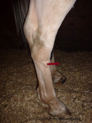 The red arrow indicates the swelling. 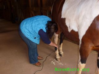 Trimming hair from swollen area 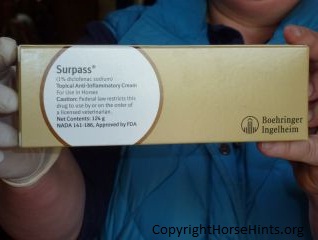 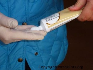 Using Surpass, a non-steroidal, anti-inflammatory (NSAID). In people speak it is known as Third Week We have stopped the Surpass. We want to see if the leg swells more. A Patchy will be confined to stall rest one more week. We drive 2 hours every day to hand walk him along with Rusty. It would be ideal if we could do this two times a day but we cannot do that given the distance. Rusty is baby sitting while A Patchy is confined to stall rest. The two of them have been together for about 6 years, now. We are hand walking the two horses in a large ring. After their 20 minute walk we hand hold both of them and let them graze. It is a long haul. Our farrier, Don Roof, came to check A Patchy, too. He believes that all will be well with some passage of time. No need to rush the recovery. I do not want one step forward and two steps back. Bill and I will slowly get back to riding with the two boys. It is really important that this tendon area not be reinjured. After the third week a re-evaluation will be made. I suspect both horses will be turned out to pasture barring any unhappy changes. We cannot keep them confined forever. Every treatment has its upside and its downside. For example, it is also possible to drain the fluid from the inflamed area and inject steroids. However, that, too, has risks such as introducing infection. I would rather not do that unless it is absolutely necessary. Apparently it is not uncommon for the fluid to return anyway. What caused this? Who knows? I suspect the horses were just frisking around and A Patchy sprained the area. His conformation is good and would not necessarily predispose him to an injury like this. He is not used in competition. Bill and I enjoy the horses, at this point in our lives, by trail riding at Manassas Battlefield and off Mary Ann's property along Lake Jackson, as well. We mostly do the slow gait and running walk along with some cantering. It was just one of those things. The horses cannot be put in a padded cell! 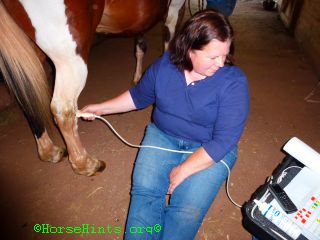  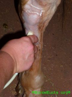 Dr. French doing ultrasound on A Patchy Tendons attach muscles and bone, and are classified as flexors (flex a joint) or extensors (extend a joint). However, some tendons will flex multiple joints and extend another (the flexor tendons of the hind limb, for example, will flex the fetlock, pastern, and coffin joint, but extend the hock joint). In this case, they are classified according to whether they flex or extend the joints of the digit. The following tendons are the main tendons found in the lower leg. When they pass over a joint, they are protected in a tendon sheath, which contains synovial fluid as a lubricant:
This is May 8, 2012 and the 6th week of A Patchy's injury. Dr. French and her vet tech, Joan, came to the barn today to give A Patchy his Lyme Disease booster. While at the barn we also discussed the progress of his tendon sheath effusion. For the past 6 weeks my husband, Bill, and I have gone to the barn every day but 3 to walk both horses for 15 minutes each day and let them graze for 15 minutes. I might add that is a round trip treck from Washington, DC to Manassas, Virginia which takes about 2 hours in travel alone. It has been extremely demanding in time and expensive; however, it is our hope that the dedication pays off. Both horses are in and have been this entire time--baby sitting each other. They are stressed when not together. Yesterday we let both of the horses out in the ring to move freely for the first time in 6 weeks. As anticipated they kicked up and were generally crazed; however, we did not see any lameness or head shaking up and down or side to side with A Patchy. Today both horses showed no sign of any further problems or recurrence of any problems after being loose in the ring. It is next to impossible to determine the time to do this. So far the decision has been good news. Today we let them out into the pasture--just the two of them--to work out their energy and graze for only two hours. This will be done for the next week, each day, if all goes well. This decision is two fold: (1) Both horses have been off the grass now for 6 weeks except for the 15 minutes of grazing each day. We do not want them to founder. (2) They have to be let out of the stalls at some point. With careful monitoring they will be watched and the timetable adjusted accordingly. Mary Ann has been wonderful through this entire episode. She really loves the horses and provides excellent care. It is our hope that Rusty will finish his Lyme Disease treatments and A Patchy will continue to heal. If all goes well Bill and I will saddle them up and ride them at a walk in the ring and on the property for several more weeks. One day at a time--then on the trail, again. Hopefully it will soon be business as usual. May 17, 2012 the 7th week - Went out to check on the horses today. They have been turned out now for several partial days--just the two of them together in a pasture. A Patchy shows no sign of any increased swelling. He looks perfectly sound in the field. Bill and I brought both horses in today, groomed them, checked them all over, then saddled them up for the first time in two months. There is a "puff" that is about 1/2" larger (when measuring the circumference of both legs in the same area) than the other hind leg where the injury occurred. That may be with him for the remainder--time will tell. (Sort of like a wind puff) We walked them to the ring, mounted and just walked around the ring. Both boys were perfect gentleman. A Patchy's footfalls felt OK. I did not feel any slippage or short stepping. His head movement was much better. It is hard to know if the insects may cause an upward movement of his head or if he may occasionally be feeling a twinge. In any event, he is much improved. We just walked around the ring in both directions, did some serpentines, walked around barrels and over cavaletti polls--boring but necessary! May 30, 2012 - Bill and I rode the horses today in the woods and along Lake Jackson. We just slow-gaited as we have had lots of rain and the footing was slippery. A Patchy was wanting to move along - forward moving - no complaints! He was not pleased that I kept him to a slow-gait. I could not detect any lameness either from his gait (footfalls) or his head movement. I believe that he is definitely on the mend. Bill and I will increase our ride time in increments. Again, I measured the leg and did a comparison to his undamaged leg. The swelling is almost completely down. The remaining "puffs" will probably be there for the duration. I just refer to it a jewelry! Both Rusty and a Patchy are turned out at night for 12 hours together--just the two of them--not with a herd. They are in by day. So far so good. Sept. 16, 2012 - A Patchy is completely sound--no tendon problems--no stifle problems. There is still some fluid that will probably be with him for the rest of his life, however, it is what we call "jewelry." It is just a blemish that does not cause any harm. I decided to not have that fluid drawn off because studies indicate that it often comes back and why should I take the risk of introducing any bacteria into the area. I do not care if there is a blemish. I have many blemishes of my own! Oct. 13, 2012 - A Patchy is doing well. Bill and I have been trail riding and have worked up to 2 hours out. In that time we walk, slow gait, fast gait and canter. Of course, we are mindful of slick footing, etc. A Patchy is moving correctly including his head carriage. There are no gait irregularities. There is no tenderness in the muscles in his back or anywhere else on his body. The only remaining sign of the injury is fluid that can be seen at the source of the original injur (puffiness). No worries! Note: "A common cause of bilateral hindlimb edema (fluid outside the joint or tendon sheath) is Lymphangitis. Inflammation of the lymphatics with restriction of lymph flow. This type of edema tend to occur from the pastern through the hock, occasionally up to the stifle in severe cases. Typically results from an unknown cause. Mild forms don't show lameness, more severe forms do as more fluid = more distention = decreased range of motion as the skin becomes more tight. Typically exercise does not resolve this type of edema. Ways we tend to treat the edema associated with lymphangitis is cold hosing the limb 10 minutes twice daily, support bandages, anti-inflammatories, and rest. Some horses may also respond to low dose steroids like dexamethazone, or naquazone (dexamethazone + trichlormethiazide). Often times multiple doses of steroids are required. We can also use sweat wraps of nitrofuracin and DMSO to help remove the edema. Often times antibiotics are ineffective."
Just Answer.HorseVeterinary Note: "...One important consideration for PRP optimization involves the concentration of white blood cells (WBCs). Although WBCs are often included because of their anti-microbial properties, which are believed to improve healing in infected wounds (one common use of PRP), PRP is often used to treat uninfected tendons or joints. Thus, the most recent data on PRP generated by Fortier's laboratory indicates that PRP low in WBCs is more beneficial in tendon healing. At present, the commercial growth of PRP products exceeds substantial research. Horse owners are encouraged with work with a veterinarian experienced in PRP when using this therapy." Optimize Platelet-Rich Plasma for Best Results Note: "Older horses can be at risk of sustaining an uncommon injury: acute rupture of the proximal (upper) superficial digital flexor tendon (SDFT) where the cannon bone meets the carpus (knee). This is because as horses age, the SDFT stiffens and becomes less elastic, decreasing its resistance to cyclic loading to the point that it can potentially tear...." Superficial Digital Flexor Tendon Rupture in Older Horses For More Information: The Conservative Approach for Healing HorsesUltrasonography of the Equine Tarsus Leg Problems Most Prevalent in Horses Horse Tendons and Ligaments |
